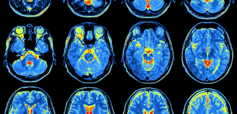
The activation of the motor cortex, a region of the brain responsible for coordinating movement, is dysfunctional in patients with fibromyalgia, a study shows.
The study, titled “Motor Cortex Function in Fibromyalgia: A Study by Functional Near-Infrared Spectroscopy,” was published in the journal Pain Research and Treatment.
Patients with fibromyalgia (FM) experience widespread muscle pain, fatigue, sleep disturbance, cognitive impairment, and other physical and psychological symptoms.
While the cause of FM remains unclear, most experimental evidence suggests that it may be due to a central failure in pain regulation and an abnormal response to pain stimuli.
There is some evidence that motor function is also impacted in FM. Studies have shown that patients with chronic pain experience changes in motor performance, and reduced motor coordination and force.
However, definitive evidence is lacking about how the motor cortex functions in FM patients compared with healthy subjects.
Measuring brain hemodynamic activity — blood flow and metabolic changes in brain tissue — during the performance of a motor task is considered a good measure of motor cortex activation. One way to measure this activity is through the use of Functional Near-Infrared Spectroscopy (fNIRS), a noninvasive technique that allows a real time detection of blood flow and metabolism changes in tissue.
In the study, researchers used fNIRS to study the hemodynamic status of the motor cortex in FM patients, and compared the results with those from healthy subjects.
The motor task that is often used in this type of neuroimaging study is known as the finger-tapping task, which consists of pressing a button with the thumb of the right hand.
Researchers evaluated two different types of motor tasks, a finger-tapping task at fixed time points (slow movement condition) and a finger-tapping task at maximum individual speed (fast movement condition).
They studied the brain hemodynamic activity in 24 FM patients and 24 healthy subjects during three different points: at rest, during slow movement, and during fast movement.
First, researchers showed that motor speed was significantly slower in FM patients compared with controls during the finger-tapping task.
During the resting state and slow movement condition, the brain hemodynamic activity of the motor cortex was similar between the two groups. However, during the fast movement task, the brain hemodynamic activity was much lower in FM patients compared with the control group.
Furthermore, this abnormality in hemodynamic activity was found to be independent of FM severity and duration. This suggests that the FM patients’ blood flow to the motor cortex of their brains is impaired during fast movement activity, and is likely the reason for their slower motor speed compared with control subjects.
“The activation of motor cortex areas is dysfunctional in FM patients, thus supporting the rationale for the therapeutic role of motor cortex modulation in this disabling disorder,” the authors concluded.

