
 Fibromyalgia syndrome (FMS) is a chronic illness that presents with immense physical pain of indeterminate cause that is sometimes accompanied by debilitating fatigue. Fibromyalgia is a syndrome — a constellation of signs and symptoms observed in, and characteristic of, a single condition — and not a specific disease with clearly defined causes and treatment. FMS is typically characterized by musculoskeletal pain that can range through the entire body, frequently accompanied by fatigue, sleep disorders, and sometimes memory and mood issues (“brain fade”). The currently prevailing theory of causation is that fibromyalgia amplifies painful sensations by affecting how their brain processes pain signals.
When a person touches a hot object or steps on something sharp, a sensation of pain is the normal response. However for sufferers from chronic pain disorders such as fibromyalgia and phantom limb pain, and even gentle skin contact can result in agonizing pain response.
A research team led by scientists at the Salk Institute in La Jolla, California, and Harvard Medical School in Cambridge, Massachusetts reports what they categorize as major breakthrough, having identified a neural mechanism in the spinal cord that apparently has the capacity to send the brain erroneous pain signals — a finding based on charting pain signaling spinal circuits in mice.
The results are reported in the journal Cell in a paper entitled: “Identification of Spinal Circuits Transmitting and Gating Mechanical Pain” (Volume 159, Issue 6, p14171432, 4 December 2014 DOI: http://dx.doi.org/10.1016/j.cell.2014.11.003), coauthored by joint first authors Bo Duan and Longzhen Cheng with Xiangyu Ren, Michael Krashes, Wendy Knowlton and Bradford B. Lowell — all of Harvard Medical School; Steeve Bourane, Olivier Britz, Christopher Padilla, Lidia Garcia-Campmany and Tomoko Velasquez of Salk Institute; Sarah E. Ross of The University of Pittsburgh; and Yun Wang of Fudan University, China, who say that their findings in murine models lay the groundwork for identifying new methodologies to treat pain disorders that have no clearly definable physical cause.
Scientists have long theorized that pain signals are sent from sensory neurons in limbs and other extremities to transmission neurons in the spinal cord, which then relay the information to the brain. The researchers note that pain information processing in the spinal cord has been long theorized to be dependent on nociceptive transmission neurons in the brain receiving input from nociceptors and mechanoreceptors — sensory neurons in the limbs and other extremities — with such inputs gated through feed-forward activation of spinal inhibitory neurons.
However, in their study, the Salk and Harvard scientists used what they call “intersectional genetic manipulations” to identify critical pain transduction components, and have been able by marking and ablating (dissipating by application of heat) six populations of spinal excitatory and inhibitory neurons, coupled with behavioral and electrophysiological analysis, were able to demonstrate that excitatory neurons expressing somatostatin include T-type cells, whose ablation causes loss of mechanical pain. They observe that inhibitory neurons marked by the expression of dynorphin represent spinal inhibitory neurons, which are necessary to gate A fibers from activating somatostatin neurons to evoke pain. At any of three steps — the extremities, the spinal cord and the brain itself they observe that pain information can be altered or even blocked before being relayed onward through the nervous system to the brain. The Spinal cord circuitry is especially key since it has the capacity to gate painful stimuli, functioning as a checkpoint between body and brain to ensure that only normal pain signals get transmitted.
 “Until now, the spinal cord circuitry involved in processing pain has remained a black box, says Dr. Martyn Goulding, Salk professor in the institution’s Molecular Neurobiology Laboratory and a co-senior author of the Cell paper in a Salk Institute release. “Identifying the neurons that make up these circuits is the first step in understanding how chronic pain stems from dysfunctional neural processing.”
The Goulding lab at Salk’s primary long-term interest and research focus are on development and functioning of neuronal circuits in the spinal cords of mammals. The lab’s research is centered on spinal circuits that control locomotion and movement, protective reflexes and posture that figure importantly in movement disorders such as spinal cord injury and neurological illness like Parkinson’s Disease. These circuits also provide neuroscientists with an ideal model system around which to structure exploration of how the nervous system generates complex behaviors — a process they refer to as the “spinal brain”. Studies at the Goulding lab employ cutting edge genetic techniques, coupled with electrophysiology, circuit mapping, imaging and behavior to investigate how the body’s motor system functions and how the spinal brain transforms sensory signals into discrete motor behaviors.
The scientists note that people afflicted with fibromyalgia syndrome or other forms of chronic pain are typically sensitive to physical stimuli that don’t normally result in pain, such as a light hand contact or a subtle changes in skin temperature. Generally referred to generally as forms of allodynia — pain due to a stimulus which does not normally provoke pain — are conditions that include fibromyalgia and nerve damage that may also be caused by diseases like diabetes, cancer and a range of autoimmune disorders.
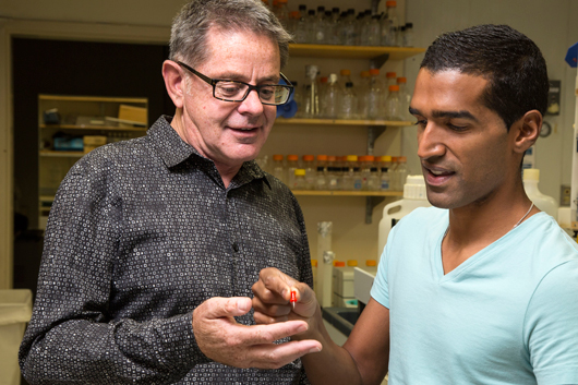 Photo Caption: Salk Professor Martyn Goulding and Jovanny Bourane, Salk research associate – Image courtesy of the Salk Institute for Biological Studies
Mysterious pain also can manifest after a limb amputation, characterized by discomfort experienced as emanating from the location where the missing appendage would be. “Phantom limb pain often subsides gradually following the amputation, but may also linger indefinitely becoming a long-term chronic pain condition for the sufferer.
 “These disorders are extremely frustrating for patients, because there is still no effective treatment for such chronic pain disorders,” says Harvard Medical School professor of neurobiology Qiufu Ma, another senior coauthor of the Cell paper. Dr. Ma’s study focus is on neural circuits involved with the senses of pain, itch, touch, and temperature. He notes that the perceived emergency of a specific sensation in response to a particular sensory stimulus is often involved with a cross talk among these labeled lines, due to polymodal nature of most sensory neurons. For example, Dr. Mac points tp the response of both pain-related and itch-related sensory neurons response to capsaicin — the chemical component in hot chili peppers that makes them “hot” in a spicy sense, noting that capsaicin injection in humans evokes only pain, due to a pain’s dominant suppression of itch. The Ma lab’s research focuses on two related areas; 1) to understand the genetic programs that control the assembly of specific sensory-labeled lines, and 2) to characterize neural circuits that underly cross interaction among distinct sensory-labeled lines by developing new genetic tools to mark and ablate specific neuronal populations in the dorsal spinal cord.
Previous research had determined the involvement of two sensory neuron types apparently involved in the spinal circuitry: pain receptors and touch receptors, and in this new study, the Salk and Harvard scientists’ objective was to identify precisely the spinal neurons active in these circuits. To that end they investigated and analyzed the roles played by the two neuronal cell types in processing pain signals in the dorsal horn, the point where sensory neurons connect with the spinal cord.
The researchers determined that dermal mechanoreceptors whose function is to detect painful mechanical stimuli are a feedback circuit component where excitatory neurons that produce somatostatin hormone are regulated by neurons that synthesize dynorphin (a natural molecular analgesic with effects similar to opiate painkilling drugs). Inhibitory neurons identified by the Salk and Harvard researchers appear to control the process whereby touch activates excitatory neurons to send pain signals to the brain — a finding that partially explains how light touch stimuli can cause substantial discomfort in persons with allodynia, ergo: if something in the pain circuitry goes haywire, so to speak, sensations of touch that normally travel through the mechanoreceptors may instead dysfunctionally activate other neurons that trigger pain signals. It is also theorized that mechanoreceptor fibers that project to the spinal cord from a missing limb may be the origin of erroneous pain stimuli.
“Normally, only pain receptors are involved in sending pain signals to the brain, but when the spinal dynorphin inhibitory neurons are lost, touch sensation are now perceived as painful,” says Dr. Goulding, who is also current holder of Salk’s Frederick W. and Joanna J. Mitchell Chair. “This really opens the door to understanding what’s happening in these pain disorders where the cause of the pain is seemingly innocuous or not known. It could be that something has gone awry in how this spinal circuitry is operating, so sensations become jumbled together and emerge as pain.”
This research was also supported by the National Institutes of Health and the Natural Science Foundation of China.
Sources:
The Salk Institute for Biological Studies
Cell
Image Credits
The Salk Institute for Biological Studies